By Astrid Bina – January 2nd 2020
Jia Xin and I are Denison Scholars working to digitise key educational Haswell Museum specimens over the summer break using a range of technologies. We are supervised by A/Prof Rosanne Quinnell. On December 17th of 2019, we were lucky to have some Haswell specimens (and some flowers) CT scanned on Cumberland Campus. We would like to thank the two radiologists there: Peter Kench and Will Rae, for giving us this wonderful opportunity. They provided us with new knowledge on how CT scans work.
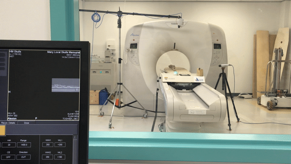
Normally, CT scans are taken from many x-rays of a patient from different angles. They are then combined to form a 3D cross-sectional image, allowing one to observe the internal organs of a patient. CT scans are commonly used for medical imaging.
CT scanning is one intriguing technique to produce 3D objects of specimens. Unlike regular photogrammetry, which only allows one to view the exterior surfaces of an object, one can see the insides of an object through CT scans. This could provide us with new information – ones that we would not get from taking individual images alone.
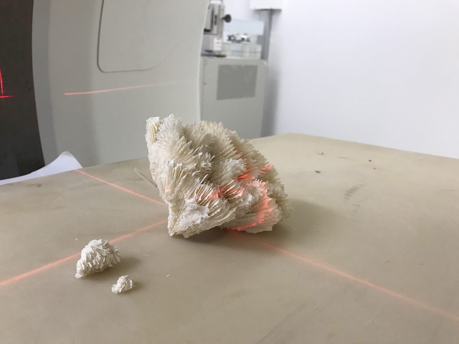
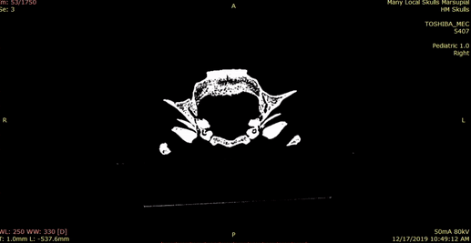
However, our peculiar request was proven to be quite a challenge, considering that the CT scanner at the Cumberland campus is typically only used for human patients. We presented an interesting set of specimens: the flowers (of course), an adult platypus in a jar, a set of marsupial skulls (kangaroo, koala and wombat), an echidna, a sea sponge, and even a small shark!
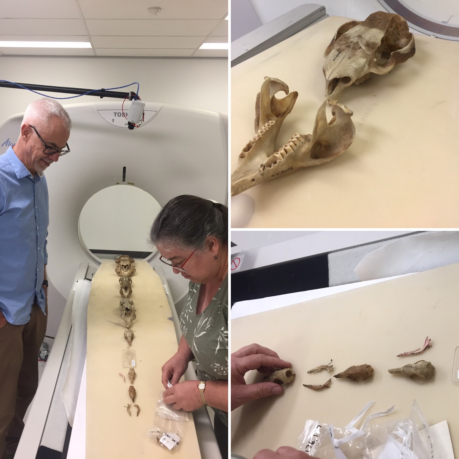
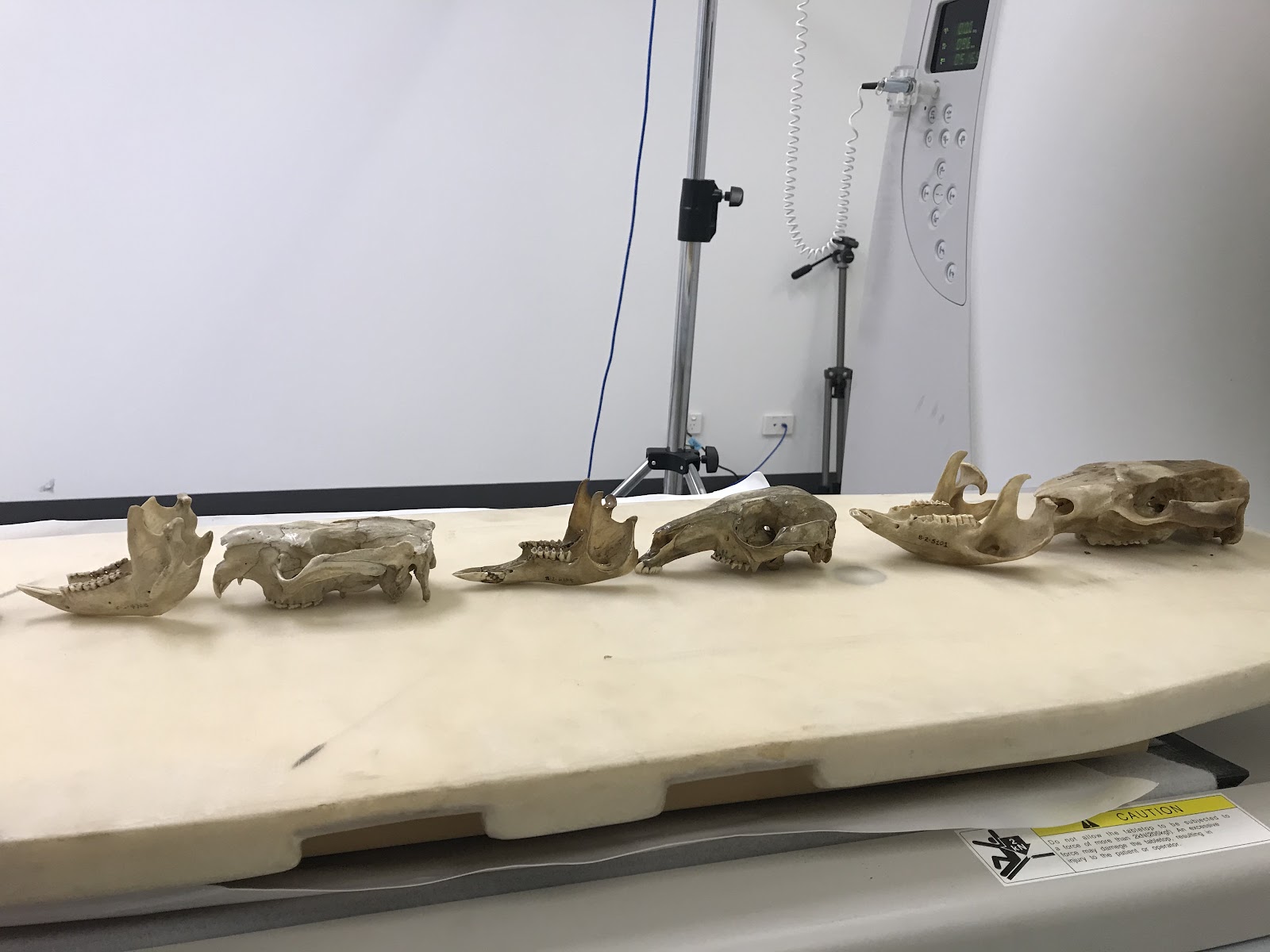
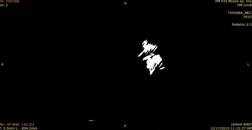
Some of the specimens we brought in store were quite easy to visualize; the skulls specifically gave some amazing output. Other specimens – especially wet specimens stored in jars like the platypus and shark, were not as easy to obtain clear CT scans from. Thankfully, Will was able to safely tweak the machine’s settings (like the energy of the radiation) to obtain clearer images. Since the specimens are non-living, higher radiation intensities could be applied. However, we still did encounter a very interesting case, where the CT scanning machine could not “detect” the shark in the jar it is stored in. This was because sharks do not have bones like we do; they are cartilaginous fish. Cartilage and water have very similar density (https://itis.swiss/virtual-population/tissue-properties/database/density/), and thus the shark, being a wet specimen, was not visible in the scans!
Overall, the scans we got from that day were very, very promising! Especially since we are still at the beginning of this journey, we still have lots of room to experiment on!
We hope that this not-so-conventional, yet very exciting technique could aid us in specimen digitisation. Together with photogrammetry, hopefully the final products can provide some fun and useful knowledge!
It would be awesome if we could CT scan that crocodile someday…